Molecular Breast Imaging (MBI)
Molecular Breast Imaging, or MBI, is the latest imaging technology we have to aid us in the diagnosis of breast cancer. It utilizes radioactive tracer (technium 99 sestambi) injected into the patient. This radiotracer localizes to the mitochondria (the “powerhouse”) of a cell which allows us to identify lesions that are metabolically active, such as breast cancer. As such, its a way to assess for lesion physiology and is another functional imaging tool.
Given that there is no special patient preparation or fasting required, it can be part of a diagnostic workup and can compliment the anatomic imaging performed the same day. The exam is performed with very light compression and can be performed quickly. In our experience it is very well tolerated by patients.
Although the exam does require the intravenous injection of radioactive tracer, we’ve managed to decrease the injected dose to less than half of what many centers use (~ 4mCu), and without any compromise in patient safety or image quality.
We feel this exciting new test best fits as a supplemental screening exam for those patients who are intermediate risk, or who are highrisk but cannot get an MRI.
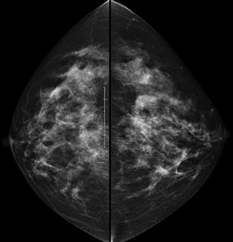
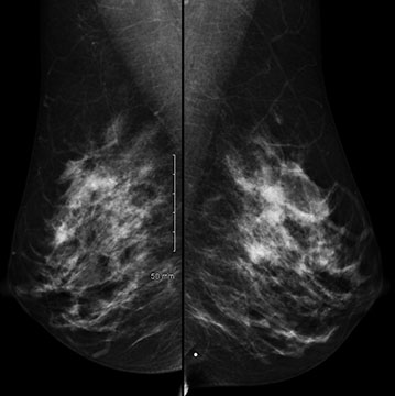
Below is a case where MBI helped to diagnose an unsuspected cancer. Patient was in her 40’s and had a normal routine screening mammogram:
 |
 |
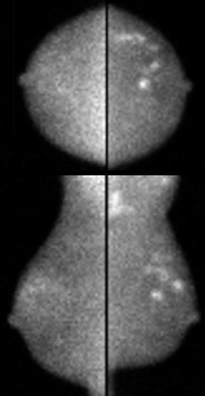
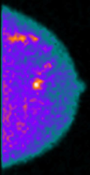
She then underwent subsequent screening MBI which demonstrated an abnormality in the left breast:
 |
 |
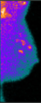 |
The MBI shows 2 separate abnormalities in the left breast, both of with came back positive for malignancy. Even knowing where the lesions are, the findings are not seen on mammography.
In summary, we are very excited about this new technology and feel privileged to bring it to our community.
links:

