

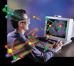
Volume-Rendered
3D Helical CT
Boca Raton Regional Hospital is one of the first facilities in the country able to offer real-time 3D volume rendering, an exciting new technology enabling unparalleled imaging of complex data sets and anatomic information.
What is 3D Helical CT?
- Displays 3D spatial relationships in
a two-dimensional image
- Volume CT data set
- Sophisticated computer post-processing
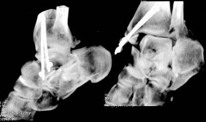
Real-time 3D CT of
Pilon fracture of distal tibia
Role of
Helical CT in Orthopedic Imaging
What is 3D Helical CT?
|
|
Role of
Helical CT in Orthopedic Imaging
CT Hardware
Requirements
|
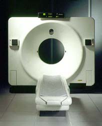 |
What is
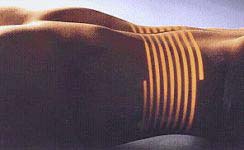
Helical (Spiral) CT?
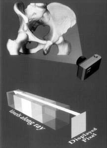
- X-ray production
- Simultaneous table & tube movement
- Produces volume data set
- Subsecond spiral acquisition- minimizes
motion artifact
- Anisotropic voxels (longer in the
z axis than in the transverse plane)
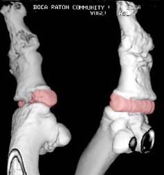
Surface Rendered 3D image
s/p 1st MPJ replacement arthroplasty utilizing a Swanson Silastic
Total Hinge Joint implant
|
|
| Surface Rendered 3D image s/p 1st MPJ replacement arthroplasty utilizing a Swanson Silastic Total Hinge Joint implant | |
3D Surface
vs Volume-Rendering
<link
to source Radiographics article (Pretorius & Fishman)>
Shaded Surface Rendering
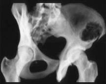
Shaded
Volume Rendering
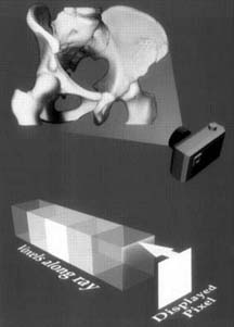
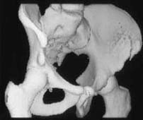
- Shaded surface rendering algorithms
- only uses first defined voxel as the surface of the bone
- Displays gross 3D relationships
- Fails to display lesions hidden
beneath the bone surface
- Tend to demonstrate stair-step artifacts.
- Uses entire data set
- Conveys more information
- Contributions of each data set voxel
are summed
- May be viewed in any plane or projection
and in a range of opacity from transparent to opaque
|
|
|
|
|
|
|
|
|
|
Computer
Hardware Requirements
- Real-time interactive display
- Eliminates need for editing prior
to 3D rendering
- Rapidly obtains clinically useful
images
- Expedites communication of results
- Anisotropic voxels interpolated
to create cuboid isotropic voxels
- Isotropic data mapped into appropriate
plane
- Rendering algorithm into 3D volume
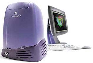
- Requires considerable computer power
(Silicon Graphics Workstation)
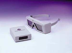
Stereo 3D
Display
|
|
Stereo 3D Display
 |
 |
3D Volume
CT may alter management
- Those who might otherwise have received conservative treatment but become surgical candidates
- Those in whom urgent surgery is delayed in favor of later, definitive arthrodesis or arthroplasty
Complex
Fractures
- Talar and calcaneal injuries easily evaluated
- 3D mapping for preoperative planning
- Complex intra-articular fractures of the
distal tibia
- Immediate surgery vs later, definitive
arthroplasty
Real-time 3D CT of Calcaneus
fracture
|
Post-operative
Imaging
|
CT and MR imaging limited due to artifact from implanted hardware Spiral CT with volume rendering able to compensate for streak artifact despite the presence of metal plates, pins, or prostheses. |
 |
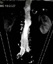
Other Applications
- Endovascular Aortic
Stent Graft used for perctaneous repair of abdomial aortic aneurysm
|
|