

Conventional
(X-ray) Mammography Cases
Conventional (X-ray) Mammography Cases
CASE 1
CLINICAL SITUATION:
The patient is a 52 year old woman found on screening mammography to have a 9 mm solid nodule in the inferior aspect of the left breast.
FINDINGS:
Mobile screening mammography ![]() , 90 degree, spot
compression imaging
, 90 degree, spot
compression imaging ![]() and ultrasound of the left breast
was performed. These films demonstrated a 9 mm solid nodule in the 5 o'clock
position of the left breast. The ultrasound
and ultrasound of the left breast
was performed. These films demonstrated a 9 mm solid nodule in the 5 o'clock
position of the left breast. The ultrasound ![]() showed
the lesion to be oval, hypoechoic and homogeneous, suggesting a benign lesion.
BIRADS category 4.
showed
the lesion to be oval, hypoechoic and homogeneous, suggesting a benign lesion.
BIRADS category 4.
PROCEDURE:
The patient was referred for percutaneous biopsy of the lesion. A hand held
Mammotome biopsy was performed. Several specimen were obtained. As it appeared
that the lesion was removed (arrows), a localization clip was left at the end
of the procedure. A two view mammogram was performed post-procedure demonstrating
removal of the lesion and excellent positioning of the clip (arrowhead) with
minimal changes at the biopsy site.![]()
PATHOLOGY:
Pathologic diagnosis revealed fibroadenoma.
DISCUSSION:
One of the reasons to use the Mammotome in a case such as this is to have the capabilities to remove a lesion. This makes interpretation of subsequent mammograms easier. Patients are generally reassured knowing the lesion is gone especially after watching the procedure on the ultrasound screen. In the past, this was typically performed by needle localized excision, a procedure which is significantly more expensive and traumatic to the patient and can result in significant deformity on subsequent mammograms.
<click here to download a video of an US guided breast biopsy (.wmv file; 33 MB)>
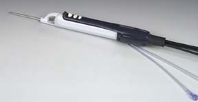
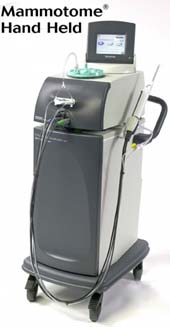
MAMMOTOME® HAND HELD
LESION REMOVAL
MAMMOTOME® HAND HELD
LESION REMOVAL
CASE 2
CLINICAL SITUATION:
The patient is a 72 year old woman with a palpable mass in the upper outer aspect of the right breast referred for diagnostic mammogram and ultrasound.
FINDINGS:
A three view mammogram showed a new ovoid 2 cm, well circumscribed mass at
the site of the palpable abnormality![]() . An ultrasound showed a complex
mass in the region measuring 1.5 x 1.0 cm in size
. An ultrasound showed a complex
mass in the region measuring 1.5 x 1.0 cm in size![]() . There
was both a hypoechoic and a hyperechoic component to the mass, which was thought
to probably be an intracystic papillary carcinoma by the interpreting physician.
The lesion was considered BIRADS Category 4.
. There
was both a hypoechoic and a hyperechoic component to the mass, which was thought
to probably be an intracystic papillary carcinoma by the interpreting physician.
The lesion was considered BIRADS Category 4.
PROCEDURE:
The Mammotome Hand Held was used to biopsy the lesion. A clip was placed as
it appeared that the majority of the lesion had been removed. Repeat two view
mammogram showed the clip in excellent position ![]() with
no residual nodule mammographically evident.
with
no residual nodule mammographically evident.
DISCUSSION:
Lesions can be removed with the 11 gauge Mammotome. A very soft lesion was found and fell into the biopsy chamber well in this case. Only eight specimens were required for lesion removal.
PATHOLOGY:
Intramammary lymph node- a diagnosis not suspected due to the mammographic appearance of the lesion, without the expected lucent center normally seen. A six month mammographic follow-up was recommended to assure stability.
<link to Breast MRI cases>
 |

|