

Rheumatoid Arthritis Screening MRI Protocol
for Wrist, Hand & Foot
by Steven Needell, M.D.
www.bocaradiology.com
Rheumatoid Arthritis Screening MRI Protocol
for Wrist, Hand & Foot
by Steven Needell, M.D.
www.bocaradiology.com
"RA" MRI Protocol:
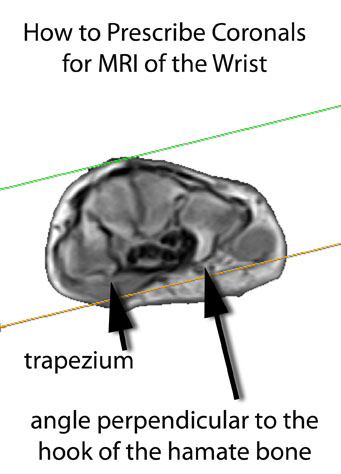
Coronal 3D T1 GRE
Coronal STIR
Axial STIR
Anatomic Coverage:
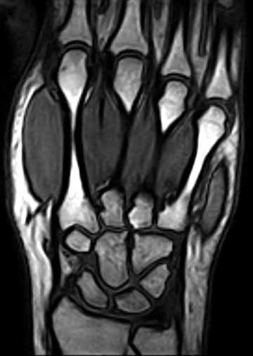
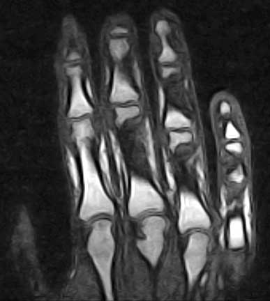
Wrist & Hand
 |
 |
WRIST Include distal radio-ulnar joint (DRUJ) through MCP joints |
HAND Include MCP joints through DIP joints |
Note: For a WRIST exam, always make sure to include the distal radio-ulnar joint (DRUJ).
If the patient has a very large hand, the MCP joints may not be able to be imaged completely.
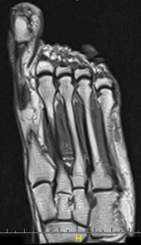
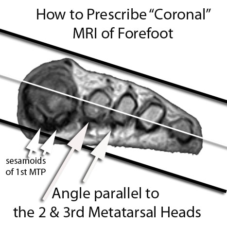
FOREFOOT
LONG AXIS "CORONAL" IMAGE OF FOREFOOT
Include Metatarsal-cuneiform joints through MTP joints
The "Coronal" plane of the forefoot here refers to the longitudinal (long axis) plane. In true anatomic position,
this is actually an axial image, but is more commonly referred to by MRI techs as "coronal" of the forefoot.
For information regarding routine image of the ankle & foot, visit: <www.bocaradiology.com/foot>