This is a case of a 61 year old male with rising PSA and two prior negative prostate biopsies, including a standard 12-core transrectal TRUS biopsy and MRI/US fused transrectal prostate biopsy most recently. Due to the location of the lesion in the anterior most aspect of the central zone, it is not surprising that the lesion was not sampled during the traditional transrectal approach. I was able to sample the lesion thoroughly utilizing MRI/US fused technology and a transperineal approach. A total of 7 core biopsy samples were taken from the target lesion, yielding Gleason 7 (3+4) disease. Of the additional 12 cores obtained, Gleason 7 (3+4) disease was also found in the apex and base, anteriorly, findings concordant with the screening MRI.
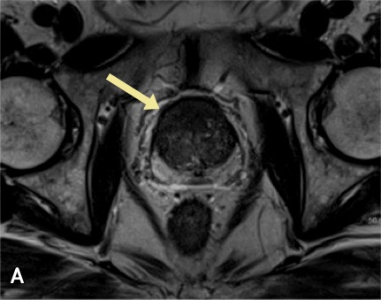
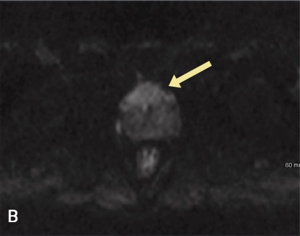
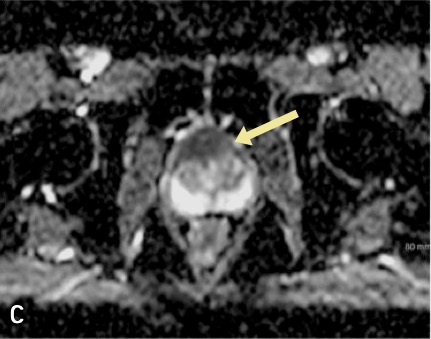
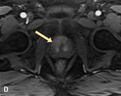
Selected axial images from the 3T, multi-parametric prostate MRI show the (A) marked T2 hypointesity (darkness) in the anterior aspect of the central zone (yellow arrow) and corresponding restricted diffusion with hyperintense (bright) signal on the diffusion image (B) and hypointense (dark) signal on the ADC map (C) both in the anterior aspect of the central zone (yellow arrows). A dynamic post contrast image (D) demonstrates corollary arterial enhancement (yellow arrow), resulting in a PI-RADS 5 lesion.
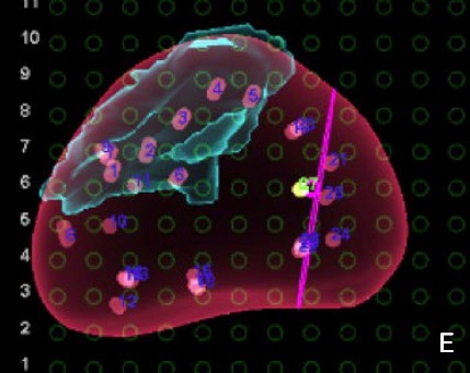
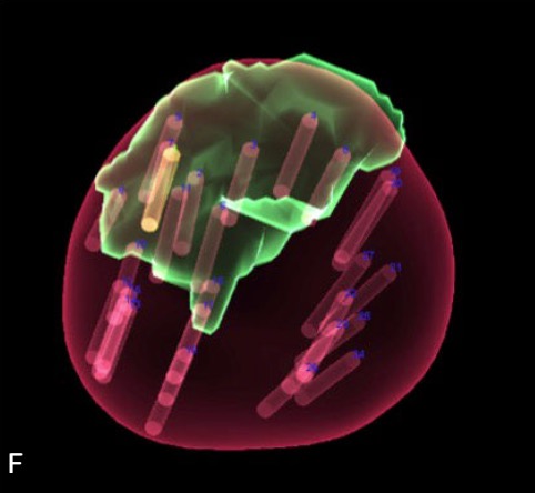
Screen captures obtained during the fused transperineal biopsy demonstrate thorough sampling of the target lesion outlined in teal (E) and green (F), the latter image representing a volume rendering of the prostate gland in 3-dimensions. Images also show the additional 12 core biopsy specimens obtained throughout the prostate gland, evenly spaced, allowing for sampling of the majority of the prostatic tissue increasing the diagnostic yield of the biopsy.
This is a case of a 76 year old male with elevated PSA and a 1.3 cm PI-RADS 5 lesion in the left posterior peripheral zone in the mid aspect of the prostate gland extending towards the apex. MRI/US fused transperineal prostate biopsy was performed, with 5 core biopsy samples taken throughout the target yielding Gleason 7 (4+3) disease with perineural invasion. Of the additional 12 cores obtained, Gleason 7 (4+3) disease was found at the left posterior base and Gleason 8 (4+4) disease at the left posterior apex and midline apex, findings concordant with the screening MRI.
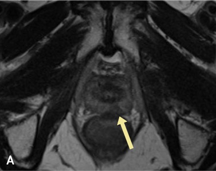
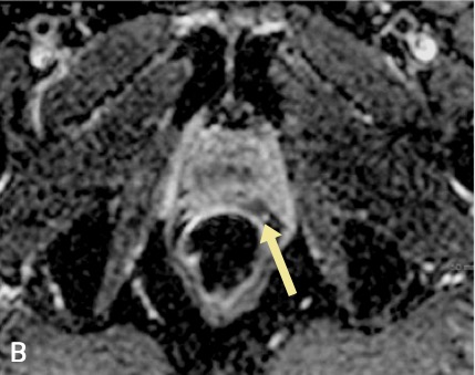
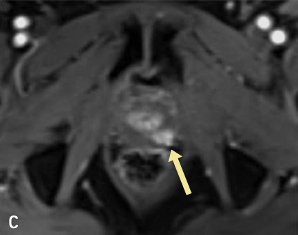
Selected axial images from the 3T, multi-parametric prostate MRI show the (A) focal T2 hypointensity (darkness) in the left posterior peripheral zone (yellow arrow) measuring approximately 1.3 cm and associated restricted diffusion with hypointense (dark) signal at the site (yellow arrow) on the ADC map (B). There is focal dynamic contrast enhancement at this site with plateau kinetics (c)
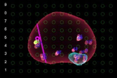
Screen captures obtained during the fused transperineal biopsy demonstrate thorough sampling of the target lesion outlined in teal, as well as additional evenly spaced core biopsy specimens obtained throughout the prostate gland.