The risk factors for heart disease include the following:
The primary cause of heart disease is the build-up of plaque (atherosclerosis) in the arteries of the heart. This build-up can cause the arteries to narrow. Plaque can also break away from the artery walls and cause a blockage. In both instances the heart muscle does not receive enough blood flow and oxygen-thus a heart attack can occur.
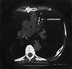
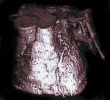
Cardiac scoring is a revolutionary, pain-free, non-invasive and inexpensive procedure requiring less than 10 minutes. Using CT imaging with sub-second scanning capability, the equipment takes 70-90 images of your coronary arteries without any injections, needles or removal of your clothing. The amount of calcium or plaque detected in your coronary arteries is used to establish your cardiac score.

First, you complete a brief risk factor questionnaire. Next, you lie down on the imaging table while a CT technologist places a few EKG leads on you. You are then asked to hold your breath while the images are taken. That's it. You can return to your regular routine.
A board certified radiologist, using high tech software, calculates your cardiac score based on the images taken.

The radiation exposure during cardiac scoring is very minimal (comparable to the radiation received during a chest x-ray). No intravenous injections or needles are required.
Not only is it safe, but it is also simple. In most cases (unless there are certain types of buttons or bra metal involved),, patients may keep their shirt or blouse on during the procedure.
Subtle early warning signs of heart disease can be detected. Cardiac scoring is the latest diagnostic tool to detect plaque in the coronary arteries. With this information, your doctor can recommend the appropriate treatment, including diet and lifestyle changes, medication and/or further testing.
Yes, the plaque build-up process can be slowed, stabilized and reversed, in some cases, through aggressive lifestyle modification and/or through medication therapies under the guidance of your physician.
|
No identifiable atherosclerotic plaque. Very low cardiovascular disease (CVD) risk |
Healthy Diet (low
in saturated fat and cholesterol) Stop Smoking Maintain Recommended Weight |
|
Minimal plaque burden Low CVD risk |
All recommendations
above PLUS Tight control of Diabetes and Hypertension Consider use of Statins in cases of High Cholesterol |
|
Mild plaque burden Moderate CVD risk |
All recommendations
above PLUS Estrogen for Post-Menopausal Women Aspirin Use Use of Statins in cases of High Cholesterol |
|
Moderate plaque burden High CVD risk |
All recommendations
above PLUS Exercise program Use of Statins in cases of high and borderline cholesterol levels Consider use of Folic Acid, Vitam E, Fish Oils |
|
Extensive plaque burden Very high CVD risk |
All recommendations above PLUS Exercise Test to rule out obstructive disease Consider angiogram for symptomatic patients or those in high risk occupations |

* Coronary Artery Calcium (CAC) Score Reference Values web tool *
McClelland RL, Chung H, Detrano R, Post W, Kronmal RA. Distribution of coronary artery calcium by race, gender, and age: results from the Multi-Ethnic Study of Atherosclerosis (MESA). Circulation. 2006;113(1):30-37.
|
|
|
|
Ideal for patients who are symptomatic |
Does not produce an image of coronary arteries |
|
Gold standard" |
Generally used only with demonstrable ischemia |
|
Direct visualization of vessel wall and lumen size; useful for angioplasty and stent placement |
Generally used only with demonstrable ischemia |
|
Noninvasive |
Does not identify stenotic lesions |
For the most current, comprehensive discussion of this state-of-the-art test and its implications, click on one of the links below:
Coronary Calcium Screening Seen Useful Beginning Between Age 40 and 50. J Am Coll Cardiol. 2005;46:807-814Coronary Artery Calcium Measurement: Agreement of Multirow Detector and Electron Beam CT. C. R. Becker, T. Kleffel, A. Crispin, A. Knez, J. Young, U. J. Schoepf, R. Haberl, and M. F. Reiser, Am. J. Roentgenol., May 1, 2001; 176(5): 1295 - 1298.
Evaluation of Subsecond Gated Helical CT for Quantification of Coronary Artery Calcium and Comparison with Electron Beam CT. J. Jeffrey Carr, et al. Am. J. Roentgenol 2000; 174:915-921.
Coronary Artery Calcification as an Indicator of Preclinical Coronary Artery Disease, by William Stanford, MD, Radiographics. 1999;19:1409-1419.