561.391.1728
by Steven Needell, M.D.
www.bocaradiology.com
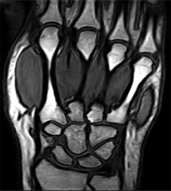
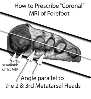
Coronal 3D T1 GRE
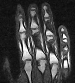
Coronal STIR
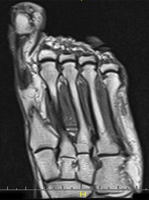
Axial STIR


ADA Compliance Policy: In accordance with the American with Disabilities Act, Boca Radiology Group strives to ensure that our web site is accessible to individuals with disabilities. If you have any questions or suggestions regarding the accessibility of this site, please contact us via email or call us at 561.391.1728, so we can ensure you receive the information you are seeking.
© 2023 All Rights Reserved