561.391.1728
from the International Society of Extremity MRI in Rheumatology
(ISEMIR) Inaugural
Conference (Chicago, IL. April 10, 2008)
Boca Radiology Group (BRG) is proud to be the nation's leader in providing interpretive telemedicine services to rheumatology practices throughout the United States. BRG offers high quality subspecialty interpretations over the internet utilizing high-speed, HIPAA- compliant teleradiology. Our professional services include supervision of MRI protocols, technologists and maintaining quality assurance. We provide prompt turnaround time of transcribed reports and immediate notification of significant findings.
MRI Techs:
Click here for technical advice on <MRI positioning and RA protocol>.
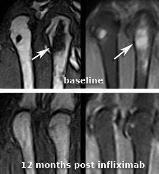
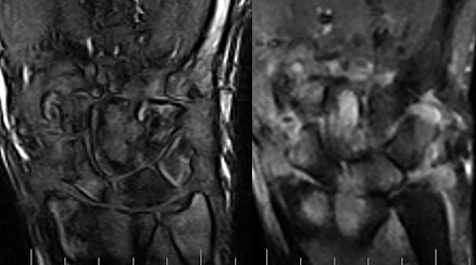
Rheumatoid arthritis (RA) affects 2.1 million people in the United States, peaking between 30-50 years of age. Early detection and treatment of rheumatoid arthritis has been shown to decrease morbidity and preserve patients' quality of life. Therapeutic medications (anti-THF therapy and conventional DMARDs) used to treat RA are quite expensive. Early identification of patients who will develop more severe, disabling disease assists our health care system in targeting which patients will benefit most from these therapies. Historically, x-ray has been considered the gold standard imaging test because of its ability to identify erosions although detection of erosions with radiographs may be delayed for up to 2 years and x-rays are unable to evaluate synovitis and osteitis in patients with aggressive disease. MRI is a sensitive modality for detection of bone marrow edema/osteitis, synovitis and erosions in RA, and has now developed an important role in therapeutic planning and assessment of treatment response. Extremity MR units using high resolution, submillimeter 3D MR imaging provide high sensitivity for early detection of disease and improved access to physicians and patients for evaluating longitudinal assessment of disease response to therapy.
ADA Compliance Policy: In accordance with the American with Disabilities Act, Boca Radiology Group strives to ensure that our web site is accessible to individuals with disabilities. If you have any questions or suggestions regarding the accessibility of this site, please contact us via email or call us at 561.391.1728, so we can ensure you receive the information you are seeking.
© 2023 All Rights Reserved